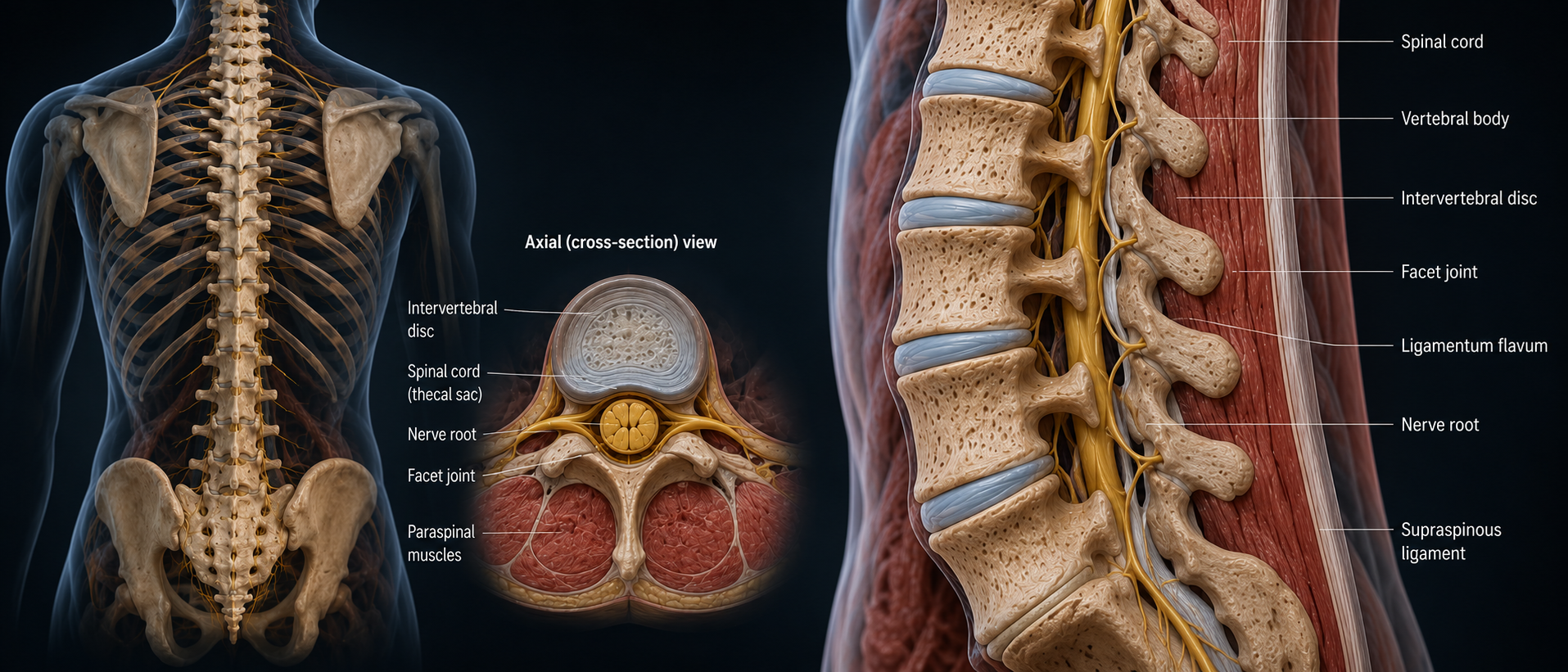
The working unit of the spine is formed by two vertebrae (blocks of bone) and the disc between them. The vertebrae connect to each other via two facet joints — arranged like roof tiles — with the disc sandwiched in between. Ligaments and the core muscles hold the unit together and allow it to move.
Disc anatomy
The disc acts as a shock absorber. In the lumbar spine, it can bear up to twice the body's weight — which is why weight reduction helps long-term disc health. Each disc has a stronger outer shell (the annulus) and a soft, jelly-like inner core (the nucleus). Think of it like a jam doughnut: the doughnut wall is the annulus, and the jam inside is the nucleus. The water content of the nucleus is what gives the disc its cushion, allowing it to absorb the stress of walking, standing, and being upright.
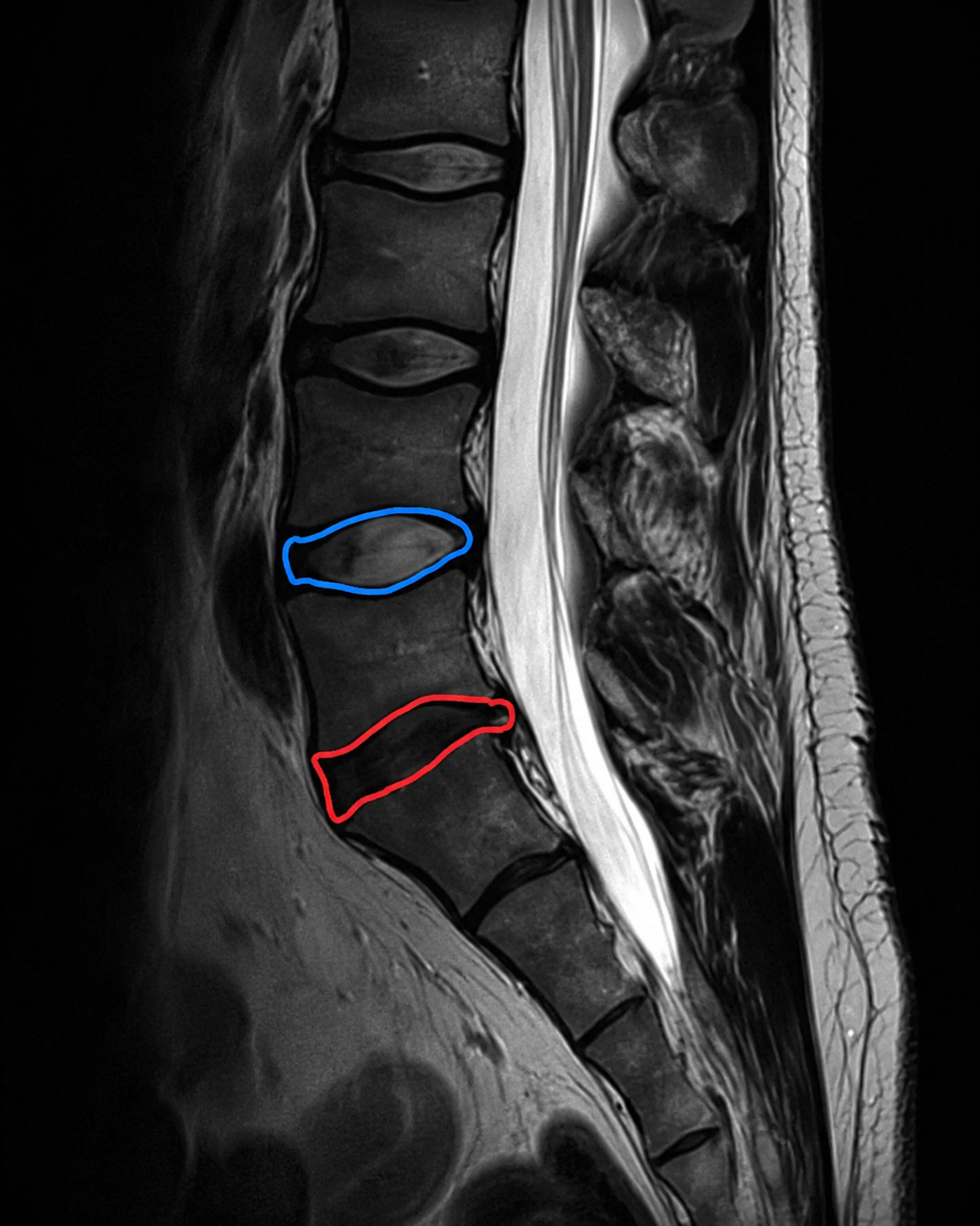
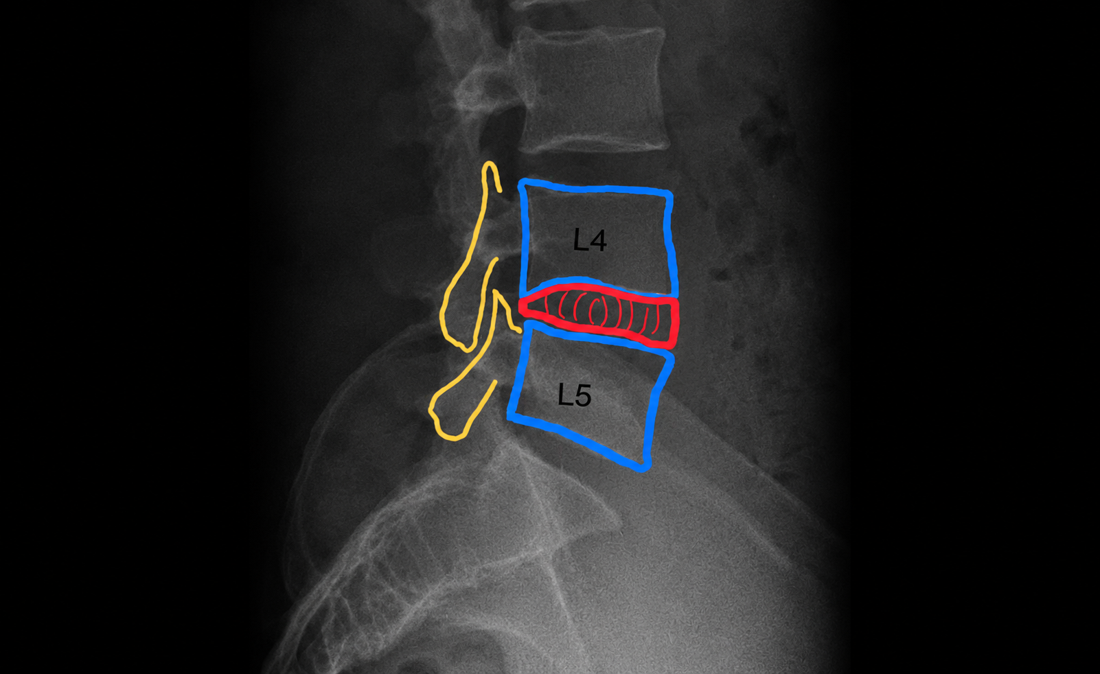
When a disc starts to lose water content, it is described as dehydrated. On MRI, a healthy disc looks pale; a dehydrated one looks dark — sometimes called a "black disc". Loss of hydration weakens the annulus and the nucleus can leak out. That escaping inner material is what produces a disc herniation, or "slipped disc".
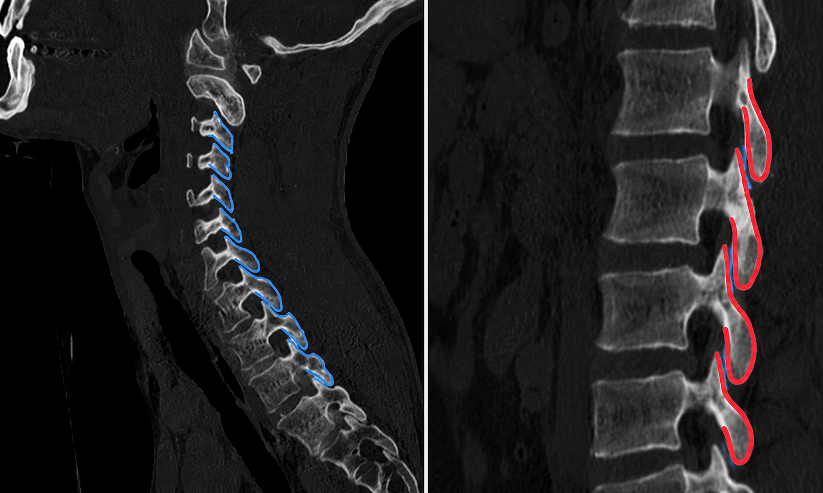
Facet joints — the roof tiles
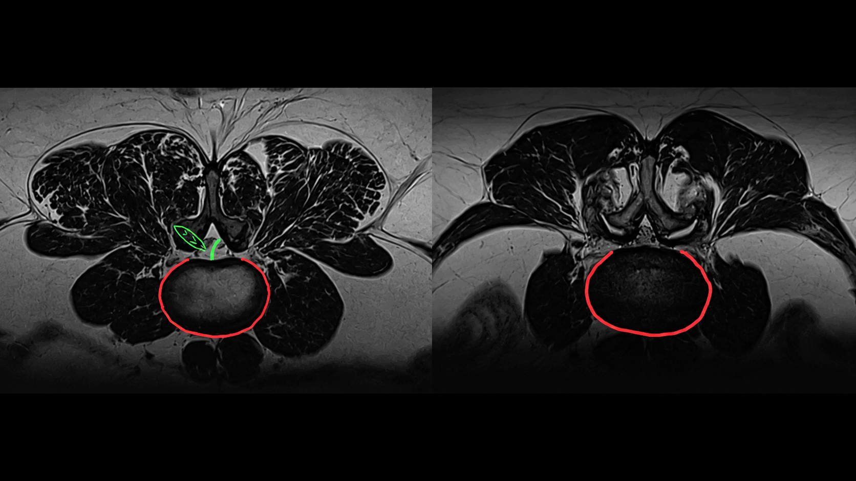
The facet joints are arranged like roof tiles, one sliding over the next with every movement. Like any other joint — hip, knee — they are lined with cartilage and lubricated by synovial fluid. They can develop arthritis, become inflamed, and they are surrounded by a tough joint capsule containing nerve endings. Those nerve endings are the target of facet denervation — burning the nerve endings — when the joint is the source of someone's back pain.
The core muscles
The core muscles keep us upright. We evolved from four-legged primates to two-legged humans, and the abdominal layers at the front, the diaphragm above, and the back muscles behind all work together to maintain that posture. As we age, posture shifts and these muscle groups — especially the back — can come under increased strain. That strain shows up as back pain and related symptoms.